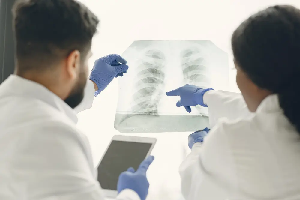
The diagnosis of lung disease relies on advanced technologies that enable physicians to visualize the air passages, as well as the adjacent anatomy. The procedures most commonly employed to evaluate patients with lung problems are bronchoscopy and endobronchial ultrasound.
Although both bronchoscopy and endobronchial ultrasound (EBUS) are minimally invasive and often performed together, the two tests have different purposes. A good understanding of the differences will help patients to be better educated and confident about their possible diagnosis and treatment. In this article, we will discuss in detail EBUS and bronchoscopy as backed by the Best bronchoscopy doctor.
What is Bronchoscopy?
Bronchoscopy is a procedure that allows physicians to visualize the internal structure of the bronchial tubes using a long flexible tube (bronchoscope).
What is Diagnosed by Bronchoscopy?
Bronchoscopy test Can Be Used To Diagnose:
- Lung COPD and other lung infections
- Blockages and/or abnormal growths in the Bronchial Tubes
- The cause of a persistent cough or hemoptysis
- Suspicious nodules or masses
In addition to visualizing inside the lung, bronchoscopy enables the physician to perform tissue collection (biopsy) of an abnormality for further evaluation.
What Is EBUS?
EBUS or Endobronchial Ultrasound uses ultrasound imaging, in addition to bronchoscopy technology, to visualize the structures around the bronchial tubes. EBUS enables the physician to visualize the tissues and lymph nodes surrounding the bronchial walls.
What is Diagnosed by EBUS?
EBUS Can Be Used To Diagnose:
- Lung cancer staging
- Enlarged lymph nodes
- Infectious disease
- Tumors that may not be directly visualized with bronchoscopy
EBUS provides physicians with extensive anatomical information without the necessity for more invasive procedures.
Key Differences between Bronchoscopy and EBUS
- Technology Used
- Bronchoscopy utilizes a camera to allow physicians to look at the inside of the airways.
- EBUS utilizes a camera, but also ultrasound technology to identify structures inside the body that are not visible through bronchoscopy.
- Area Being Examined
- With Bronchoscopy test in Mohali, the physician will only look inside the airways.
- EBUS will identify not just the structures inside the airways, but also the surrounding structures, lymph nodes, and the tissues surrounding those structures.
- Diagnostic Capability
- Bronchoscopy is used for diagnosing abnormalities within the airways that can be seen with the use of a camera.
- EBUS can identify deep-seated tumors and can be used to stage cancers better than bronchoscopy.
- Precision of Biopsy
- EBUS can perform more precise and better-targeted biopsies than bronchoscopy. This is especially true when it involves obtaining lymph node biopsies. This reduces the need for surgically obtaining lymph node biopsies in many cases.
When Should These Tests Be Done?
Your doctor may recommend a bronchoscopy or EBUS to evaluate you for:
- Persistent cough or lung symptoms that cannot be explained
- Lung abnormalities identified on imaging studies
- Suspected lung cancer
- Enlarged lymph nodes are located in the chest
Sohana Hospital Mohali provides the best solutions for patients looking for the Best pulmonologist in Chandigarh, thanks to its advanced technology and the most trusted and experienced lung specialists in the tricity. Book your appointment today!